- Transcatheter Aortic Valve Replacement
- Cardiac Catheterization (Angiogram)
- Coronary Angioplasty
- Peripheral Angioplasty
- Treatment of structural heart diseases
Transcatheter Aortic Valve Replacement
What is Aortic Stenosis?
Aortic stenosis is a condition where the aortic valve, the valve that allows blood to pass from the main pumping chamber of the heart to the rest of the body, becomes stiff and obstructed. Uncorrected, this can lead to debilitating symptoms (shortness of breath, chest pain, passing out), reduced pumping efficiency of the heart, and/or death. For patients with severe aortic stenosis who are at moderate, high or extreme risk for surgical aortic valve replacement, transcatheter aortic valve replacement (TAVR) is an effective option. What was previously major open-heart surgery is now being performed minimally invasively on an awake and talking patient with excellent results.
About Dr. Elmore
What is TAVR?
Much less invasive than traditional surgical valve replacement, TAVR is a nonsurgical procedure that allows the implantation of a new heart valve without the need to open the chest. It is performed under local anesthesia via passage of a special catheter (hollow tube) into the blood vessel in the groin. Once inserted, the catheter is advanced to the heart and the new valve is implanted. Only a tiny incision site remains, which heals on its own within a few days; without the need for stitches.
There are many significant benefits for patients who qualify for TAVR, including:
- Light sedation and local anesthesia
- Minimal blood loss compared with open valve repair
- Lower respiratory and cardiac complications
- Fewer days in intensive care
- Shorter time in the hospital
- Faster recovery
Who is eligible for TAVR?

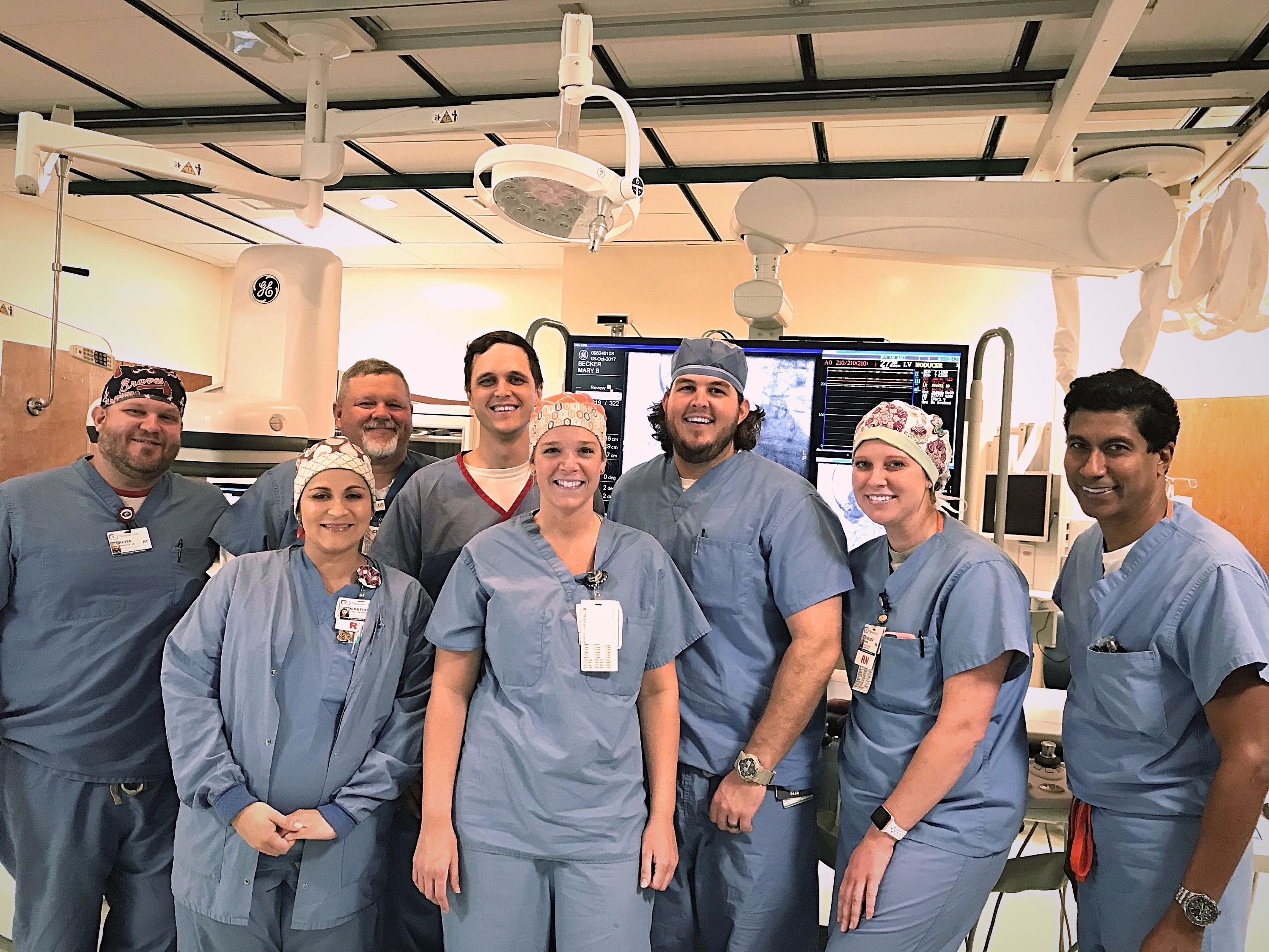
Transcatheter aortic valve replacement is indicated for patients with severe native aortic valve stenosis and patients with failed bioprosthetic surgically implanted aortic valves that are at moderate and high surgical risk. TAVR is also approved for patients who cannot undergo open heart surgery due to poor heart conditions and other health problems that make open-heart surgery too high risk (prohibitive surgical risk patients). The surgical risk is based on a number of factors, including prior surgeries, the presence of other diseases, age and your overall physical condition. Our team of experienced, highly trained, individuals, consisting of interventional cardiologists, cardiac surgeons, and specially trained nurses will evaluate you to determine your candidacy for the procedure.
What testing will you need to have to determine your eligibility?
- An echocardiogram (ultrasound) of your heart will assess heart valves and function. Your cardiologist may have already done this test. There is no preparation for this test.
- You will be scheduled for a heart catheterization to evaluate the blood flow to your heart muscle. This test will require you to have recent blood work and you may need to stop certain medications to prepare. The night prior to the test you should not have any food or drink.
- You will be scheduled for a special computed tomography (CT) scan to assess your heart valve and also evaluate the condition and size of the blood vessels that will be used to insert the valve. Our team will schedule this test and you may need to have blood work prior to scheduling this test.
- There will be periodic blood work done.
- There may be additional tests recommended.
What are the outcomes for TAVR?
Our TAVR team uses a minimalist approach to TAVR, which enables us to perform the procedure under local anesthesia and minimum sedation. This approach ultimately benefits patients, reducing their length of stay at the hospital and potential complications related to general anesthesia, while maintaining an equivalently high standard of efficacy. TAVR has shown to be markedly superior to medical management in ineligible surgical patients, and equivalent or better than open-heart surgery in high-risk surgical patients.
For moderate risk surgical patients, TAVR has been demonstrated to be equivalent to open heart surgery over a one-year follow-up period.
What to expect post procedure?
The first night you will stay in the Cardiovascular Intensive Care Unit (CVICU). Most patients stay in the hospital approximately 48 hours. However, some patients are able to go home the day after the procedure.
Our team will continue to assess your discharge/home going needs. Remember, this is not a surgical procedure: therefore you should be able to do most of your activities once back home, but will need some assistance for at least one week. Your home-going support should consist of family members or friends that can assist you continuously for approximately five days with things such as meal preparation and household duties. Once you are able to bath and dress comfortably along with basic meal preparation and getting around the house (i.e. take stairs) without assistance, your support system and care partner can allow you to be more independent.
You will return for a clinic appointment with our team at one week, 30 days and one year. You should also schedule follow-up appointments with your cardiologist and/or primary care provider. After discharge, we may facilitate cardiac rehabilitation or physical therapy appointments.
Timelines for symptom relief and degree of improvement after the procedure may vary. Some patients start feeling improvement as early as one week, and at one-month post-procedure, most patients feel significantly better. Combining physical therapy or cardiac rehabilitation as part of the recovery process may further enhance the overall improvement.
Cardiac Catheterization (Angiogram)
Cardiac Catheterization (Angiogram)
Cardiac catheterization is a medical procedure we perform to diagnose and treat certain heart conditions.
A long, thin, flexible tube called a catheter is put into a blood vessel in your arm, groin (upper thigh), or neck and threaded to your heart. Through the catheter, your doctor can perform diagnostic tests and treatments on your heart.
How to prepare for a Cardiac Catheterization
Don’t eat or drink anything for six to eight hours before the test. If you have diabetes, discuss this with your doctor. Not eating can affect your blood sugar and adjustments may need to be made to your insulin dosage.
Discuss any medicines you are taking with your doctor. He or she may want you to stop taking them before the test, especially if you are taking a blood-thinner such as Coumadin® (warfarin) or anti-platelet medicines such as aspirin or Plavix®. It is important and helpful to bring a list of your allergies, medicines and dosages to the procedure, so the healthcare team knows exactly what you are taking and how much.
It may not be safe to drive after having cardiac catheterization, so you must arrange for a ride home.
What to Expect During Cardiac Catheterization
We perform cardiac catheterizations in a hospital. During the procedure, you’ll be kept on your back and awake. This allows you to follow your doctor’s instructions during the procedure. You’ll be given medicine to help you relax, which may make you sleepy.
Your doctor will numb the area on the arm, groin or neck, where the catheter will enter your blood vessel. A needle is used to make a small hole in the blood vessel. Through this hole your doctor will put a tapered tube called a sheath.
Next, your doctor will put a thin, flexible wire through the sheath and into your blood vessel. This guide wire is then threaded through your blood vessel to your heart. The wire helps your doctor position the catheter correctly. Your doctor then puts a catheter through the sheath and slides it over the guide wire and into the coronary arteries.
Special digital images are taken of the guide wire and the catheter as they’re moved into the heart. The movies help your doctor see where to position the tip of the catheter.
When the catheter reaches the right spot, your doctor will use it to do tests or treatments on your heart. For example, your doctor may do angioplasty and stenting.
During the procedure, your doctor may put a special dye (contrast) in the catheter. This dye will flow through your bloodstream to your heart. Once the dye reaches your heart, it will make the inside of your heart’s arteries show up on an x-ray called an angiogram. This test is called coronary angiography.
Coronary angiography can show how well blood is being pumped out of the heart’s main pumping chambers, which are called ventricles. When the catheter is inside your heart, your doctor may use it to take blood samples from different parts of the heart or to do minor heart surgery.
To get a more detailed view of a blocked coronary artery, your doctor may do intracoronary ultrasound. For this test, your doctor will thread a tiny ultrasound device through the catheter and into the artery. This device gives off sound waves that bounce off the artery wall (and its blockage) to make an image of the inside of the artery.
If the angiogram or intracoronary ultrasound shows blockages or other possible problems in the heart’s arteries, your doctor may use angioplasty to open the blocked arteries.
After your doctor does all of the needed tests or treatments, he or she will pull back the catheter and take it out along with the sheath. The opening left in the blood vessel will then be closed up and bandaged. A small weight may be put on top of the bandage for a few hours to apply more pressure. This will help prevent major bleeding from the site.
What to Expect After Cardiac Catheterization
After cardiac catheterization, you will be moved to a special care area. You will rest there for several hours or overnight. During that time, your movement will be limited to avoid bleeding from the site where the catheter was inserted.
While you recover in this area, nurses will check your heart rate and blood pressure regularly. They also will check for bleeding from the catheter insertion site.
A small bruise may develop on your arm, groin or neck at the site where the catheter was inserted. That area may feel sore or tender for about a week. Let your doctor know if you develop problems such as:
A constant or large amount of bleeding at the insertion site that can’t be stopped with a small bandage
Unusual pain, swelling, redness, or other signs of infection at or near the insertion site.
Talk to your doctor about whether you should avoid certain activities, such as heavy lifting, for a short time after the procedure.
What Are the Risks of Cardiac Catheterization?
Cardiac catheterization is a common medical procedure that rarely causes serious problems. However, complications can include:
- Bleeding, infection, and pain where the catheter was inserted.
- Damage to blood vessels. Rarely, the catheter may scrape or poke a hole in a blood vessel as it’s threaded to the heart.
- An allergic reaction to the dye used.
Other, less common complications of the procedure include:
- Arrhythmias (irregular heartbeats). These often go away on their own, but may need treatment if they persist.
- Damage to the kidneys caused by the dye used.
- Blood clots that can trigger stroke, heart attack, or other serious problems.
- Low blood pressure.
- A buildup of blood or fluid in the sac that surrounds the heart. This fluid can prevent the heart from beating properly.
As with any procedure involving the heart, complications can sometimes be fatal. However, this is rare with cardiac catheterization.
Coronary Angioplasty
What Is Coronary Angioplasty?
Coronary angioplasty is a procedure we use to open blocked or narrowed coronary (heart) arteries. The procedure improves blood flow to the heart muscle.
Over time, a fatty substance called plaque can build up in your arteries, causing them to harden and narrow. This condition is called atherosclerosis.
Atherosclerosis can affect any artery in the body. When atherosclerosis affects the coronary arteries, the condition is called coronary heart disease (CHD) or coronary artery disease.
Angioplasty can restore blood flow to the heart if the coronary arteries have become narrowed or blocked because of CHD.
Angioplasty is a common medical procedure. It may be used to:
- Improve symptoms of CHD, such as angina and shortness of breath. (Angina is chest pain or discomfort.)
- Reduce damage to the heart muscle caused by a heart attack.
- Reduce the risk of death in some patients.
Angioplasty is done on more than 1 million people a year in the United States. Serious complications don’t occur often. However, they can happen no matter how careful your doctor is or how well he or she does the procedure.
How Is Coronary Angioplasty Done?
Before coronary angioplasty is done, your doctor will need to know the location and extent of the blockages in your coronary (heart) arteries. To find this information, your doctor will use coronary angiography. This test uses dye and special x-rays to show the insides of your arteries.
During angiography, a small tube called a catheter is inserted in an artery, usually in the groin (upper thigh). The catheter is threaded to the coronary arteries.
Special dye, which can be seen on an x-ray, is injected through the catheter. X-ray pictures are taken as the dye flows through your coronary arteries. This outlines blockages, if any are present, and tells your doctor the location and extent of the blockages.
For the angioplasty procedure, another catheter with a balloon on its tip (a balloon catheter) is inserted in the coronary artery and positioned in the blockage. The balloon is then expanded. This pushes the plaque against the artery wall, relieving the blockage and improving blood flow.
A small mesh tube called a stent usually is placed in the artery during angioplasty. The stent is wrapped around the deflated balloon catheter before the catheter is inserted in the artery.
When the balloon is inflated to compress the plaque, the stent expands and attaches to the artery wall. The stent supports the inner artery wall and reduces the chance of the artery becoming narrowed or blocked again.
Some stents are coated with medicines that are slowly and continuously released into the artery. These are called drug-eluting stents. The medicines help prevent the artery from becoming blocked with scar tissue that grows in the artery.
How to prepare for a Coronary Angioplasty
Don’t eat or drink anything for six to eight hours before the test. If you have diabetes, discuss this with your doctor. Not eating can affect your blood sugar and adjustments may need to be made to your insulin dosage.
Discuss any medicines you are taking with your doctor. He or she may want you to stop taking them before the test, especially if you are taking a blood-thinner such as Coumadin® (warfarin) or anti-platelet medicines such as aspirin or Plavix®. It is important helpful to bring a list of your allergies, medicines and dosages to the procedure, so the healthcare team knows exactly what you are taking and how much.
It may not be safe to drive after having cardiac catheterization, so you must arrange for a ride home.
What to Expect Before Coronary Angioplasty
We perform coronary angioplasties at a hospital. Once the angioplasty is scheduled, your doctor will advise you:
When to begin fasting (not eating or drinking) before the procedure. What medicines you should and shouldn’t take on the day of the angioplasty.
When to arrive at the hospital and where to go.
Even though angioplasty takes only 1 to 2 hours, you’ll likely need to stay in the hospital overnight or longer. Your doctor may advise you not to drive for a certain amount of time after the procedure, so you may have to arrange for a ride home.
Preparation
In the cath lab, you’ll lie on a table. An intravenous (IV) line will be placed in your arm to give you fluids and medicines. The medicines will relax you and prevent blood clots from forming.
To prepare for the procedure, the area where your doctor will insert the catheter will be shaved. The catheter usually is inserted in your groin (upper thigh). The shaved area will be cleaned and then numbed. The numbing medicine may sting as it’s going in.
The Procedure
During angioplasty, you’ll be awake but sleepy.
Your doctor will use a needle to make a small hole in an artery in your arm or groin. A thin, flexible guide wire will be inserted into the artery through the small hole. The needle is then removed, and a tapered tube called a sheath is placed over the guide wire and into the artery.
Next, your doctor will put a long, thin, flexible tube called a guiding catheter through the sheath and slide it over the guide wire. The catheter is moved to the opening of a coronary artery, and the guide wire is removed.
Next, your doctor will inject a small amount of special dye through the catheter. This will help show the inside of the coronary artery and any blockages on an x-ray picture called an angiogram.
Another guide wire is then put through the catheter into the coronary artery and threaded past the blockage. A thin catheter with a balloon on its tip (a balloon catheter) is threaded over the wire and through the guiding catheter.
The balloon catheter is positioned in the blockage. The balloon is then inflated. This pushes the plaque against the artery wall, relieving the blockage and improving blood flow through the artery. Sometimes the balloon is inflated and deflated more than once to widen the artery. Afterward, the balloon catheter, guiding catheter, and guide wire are removed.
A drill-like device called a rotablator is sometimes used to remove very hard plaque from the artery.
Your doctor may put a stent (small mesh tube) in your artery to help keep it open. If so, the stent will be wrapped around the balloon catheter.
When your doctor inflates the balloon, the stent will expand against the wall of the artery. When the balloon is deflated and pulled out of the artery with the catheter, the stent remains in place in the artery.
Peripheral Angioplasty
Peripheral Angioplasty
Angioplasty (also called percutaneous transluminal angioplasty, or PTA) is a procedure in which a thin, flexible tube called a catheter is inserted through an artery and guided to the place where the artery is narrowed.
When the tube reaches the narrowed artery, a small balloon at the end of the tube inflates for a short time. The pressure from the inflated balloon presses the fat and calcium (plaque) against the wall of the artery to improve blood flow.
In angioplasty of the aorta (the major abdominal artery) or the iliac arteries (which branch off from the aorta), a small, expandable tube called a stent is usually put in place at the same time. Reclosure (restenosis) of the artery is less likely to occur if a stent is used. Stents are less commonly used in angioplasty of smaller leg arteries like the femoral, popliteal, or tibial arteries, because they are subject to trauma and damage in these locations.
What to Expect After Treatment
After the procedure, you will rest in bed for 6 to 8 hours. You may have to stay overnight in the hospital. After you leave the hospital, you can most likely return to normal activities.
Why It Is Done
This procedure is commonly used to open narrowed arteries that supply blood flow to the heart. It may be used on short sections of narrowed arteries in people who have peripheral arterial disease (PAD).
How Well It Works
Angioplasty can restore blood flow and relieve intermittent claudication. Angioplasty can help you walk farther without leg pain than you did before the procedure.
How well angioplasty works depends on the size of the blood vessel, the length of blood vessel affected, and whether the blood vessel is completely blocked.
In general, angioplasty works best in the following types of arteries:
- Larger arteries.
- Arteries with short narrowed areas.
- Narrowed, not blocked, arteries.
Risks
Complications related to the catheter include:
- Pain, swelling, and tenderness at the catheter insertion site.
- Irritation of the vein by the catheter (superficial thrombophlebitis).
- Bleeding at the catheter site.
- A bruise where the catheter was inserted. This usually goes away in a few days.
- Serious complications are rare. These complications may include:
- Sudden closure of the artery.
- Blood clots.
- A small tear in the inner lining of the artery.
- An allergic reaction to the contrast material used to view the arteries.
- Kidney damage. In rare cases, the contrast material can damage the kidneys, possibly causing kidney failure.
Radiation risk
There is always a slight risk of damage to cells or tissues from being exposed to any radiation, including the low levels of X-ray used for this test. But the risk of damage from the X-rays is usually very low compared with the potential benefits of the test.
What to Think About
In some cases, bypass surgery may be the best treatment choice. This treatment choice depends on your risks with the procedure, the size of the arteries, and the number and length of the blockages or narrowing in the arteries.
Your doctor may recommend that you try an exercise program and medicine before he or she recommends that you have angioplasty.
Treatment of structural heart diseases: Closure of ASD and PFO holes in heart
Closure of Holes in the Heart (ASD and PFO)
The septum is the wall that separates the chambers on the left side of the heart from those on the right. The wall prevents mixing of blood between the two sides of the heart.
An atrial septal defect (ASD) is a hole in the part of the septum that separates the atria—the upper chambers of the heart. This heart defect allows oxygen-rich blood from the left atrium to flow into the right atrium instead of flowing to the left ventricle as it should. At the time of the procedure, a transesophageal echo (TEE) or intracardiac echo (ICE) may be performed to direct the physician.
Normal Heart and Heart with Atrial Septal Defect
An ASD can be small or large. Small ASDs allow only a little blood to leak from one atrium to the other. Very small ASDs don’t affect the way the heart works and don’t require any treatment. Many small ASDs close on their own as the heart grows during childhood.
Medium to large ASDs allow more blood to leak from one atrium to the other, and they’re less likely to close on their own.
Half of all ASDs close on their own or are so small that no treatment is needed. Medium to large ASDs that need treatment can be repaired using a catheter procedure or open-heart surgery.
Ventricular septal defect (VSD)
A VSD is a hole in the part of the septum that separates the ventricles—the lower chambers of the heart. The hole allows oxygen-rich blood to flow from the left ventricle into the right ventricle instead of flowing into the aorta and out to the body as it should.
Normal Heart & Heart with Ventricular Septal Defect
A VSD can be small or large. A small VSD doesn’t cause problems and may close on its own. Large VSDs cause the left side of the heart to work too hard. This increases blood pressure in the right side of the heart and the lungs because of the extra blood flow.
The increased work of the heart can cause heart failure and poor growth. If the hole isn’t closed, high blood pressure can scar the delicate arteries in the lungs. Open-heart surgery is used to repair VSDs.
Narrowed Valves
Simple congenital heart defects also can involve the heart’s valves. These valves control the flow of blood from the atria to the ventricles and from the ventricles into the two large arteries connected to the heart (the aorta and the pulmonary artery). Valves can have the following types of defects:
- Stenosis. This defect occurs if the flaps of a valve thicken, stiffen, or fuse together. This prevents the valve from fully opening. The heart has to work harder to pump blood through the valve.
- Atresia. This defect occurs if a valve doesn’t form correctly and lacks a hole for blood to pass through. Atresia of a valve generally results in more complex congenital heart disease.
- Regurgitation. This is when the valve doesn’t close completely, so blood leaks back through the valve.
The most common valve defect is called pulmonary valve stenosis, which is a narrowing of the pulmonary valve. This valve allows blood to flow from the right ventricle into the pulmonary artery. From there it flows to the lungs to pick up oxygen.
Pulmonary valve stenosis can range from mild to severe. Most children who have this defect have no signs or symptoms other than a heart murmur. (A heart murmur is an extra or unusual sound heard during a heartbeat.) Treatment isn’t needed if the stenosis is mild.
In babies who have severe pulmonary valve stenosis, the right ventricle can get very overworked trying to pump blood to the pulmonary artery. These infants may have symptoms such as rapid or heavy breathing, fatigue (tiredness), or poor feeding.
If a baby also has an ASD or patent ductus arteriosus (PDA), oxygen-poor blood can flow from the right side of the heart to the left side. This can cause cyanosis. Cyanosis is a bluish tint to the skin, lips, and fingernails. It occurs because the oxygen level in the blood leaving the heart is below normal.
Older children who have severe pulmonary valve stenosis may have symptoms such as fatigue while exercising. Severe pulmonary valve stenosis is treated with a catheter procedure.

